📖 Read more: Bioprinting Organs: Kidneys on Demand
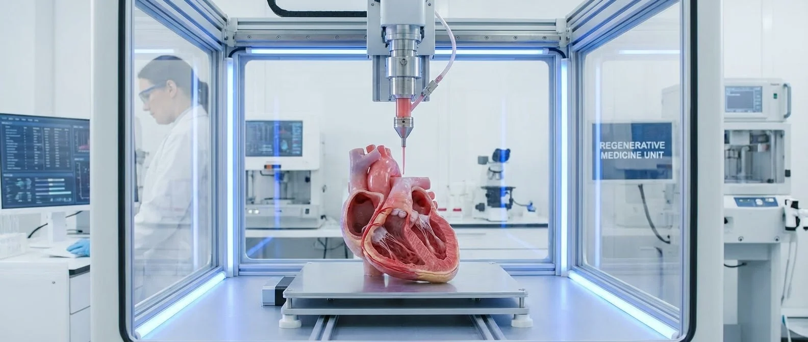
🔬 What Is 3D Organ Bioprinting?
3D bioprinting uses three-dimensional printing to combine cells, growth factors, bio-inks, and biomaterials into functional biological structures. Unlike conventional 3D printers that use plastic filament, bioprinters use “ink” made of living cells suspended in hydrogels that nourish and keep them alive.
The process follows three stages: Pre-bioprinting (tissue biopsy, CT/MRI scanning for the model), Bioprinting (layer-by-layer deposition of bio-inks), and Post-bioprinting (maturation in a bioreactor). Each stage is critical for the mechanical integrity of the final organ.
🏥 The Milestones That Changed Everything
The story of bioprinting began at Boston Children's Hospital, where Anthony Atala and his team hand-built scaffolds and layered them with patient cells to create replacement urinary bladders. Seven years after transplantation, the patients were still healthy — pushing Atala to automate the process.
⚙️ How It Works: Printing Technologies
There are three main bioprinting technologies, each with unique advantages:
Extrusion-Based
The most widespread method. Bio-ink is pushed through a nozzle layer by layer. Ideal for large structures. Uses pneumatic, piston, or screw-driven mechanisms.
Laser-Assisted
Uses laser beams to transfer cells without mechanical stress. High resolution but high cost. Best suited for fine, precise tissue structures.
Inkjet (Droplet)
Droplet printers deposit small quantities of bio-ink at precise positions. Fast and cost-effective. Suitable for proteins, nucleic acids, and small tissue structures.
📖 Read more: Organoids: Mini Human Organs in a Dish
🚀 FLOAT Technology: 50x Faster Printing
One of the biggest challenges was slow print speed — larger structures require hours, increasing the risk of cell damage. Researchers at the University at Buffalo developed the FLOAT method (Fast hydrogel stereolithography printing), using light and hydrogels.
How FLOAT Works
Light is directed into a vat of liquid hydrogel packed with living cells. Wherever the light hits, the gel solidifies. By programming precise light patterns, complex structures are created in record time. A model of a human hand was printed in just 19 minutes instead of 6 hours. The technique cuts cell damage dramatically.
🧬 Bio-Inks: The Heart of the Technology
Bio-inks are the critical ingredient. They consist of living cells mixed in a “cocktail” of enzymes and nutrients within a hydrogel. The hydrogel acts as a scaffold — allowing cells to attach, grow, and differentiate into their mature form.
Bio-ink selection determines success: extrusion systems use cell-encapsulating hydrogels, while techniques requiring cross-linking utilize GelMA (Gelatin Methacryloyl). Each tissue type requires a different formulation — hearts need elasticity, bones need hardness, cartilage needs compression resistance.
🏗️ The Challenge: Blood Vessels and Complexity
Printing fully functional organs is still the hardest part. Artificial organs — liver, kidneys, lungs — still lack critical elements:
- Blood vessels: Without a vascular network, cells deep inside the organ cannot receive oxygen and nutrients
- Tubules: Kidneys require urine-collecting tubules — an extremely difficult structure to reproduce
- Billions of cells: An adult liver contains ~100 billion cells — reproducing them in a lab requires enormous time
- Electrical conductivity: A bioprinted heart must meet not only structural but also electrical signal propagation requirements
"The technique significantly reduces part deformation and cellular injuries caused by the prolonged exposure to the environmental stresses you commonly see in conventional 3D printing methods."
📖 Read more: Xenotransplantation: Animal Organs in Humans
🔮 The Timeline: When Will We Print Organs?
Progress is rapid, but realistic estimates point to gradual adoption:
📅 2025-2028: Simple Tissues
Skin, cartilage, ear structures. Clinical trials are already underway. The first commercial products are expected soon.
📅 2028-2035: Small Organs
Thyroid, ovaries, small liver segments. Stanford aims to place a 3D-printed heart in a pig by 2028.
📅 2035-2045: Full Organs
Kidneys, liver, lungs made from the patient's own cells. The ultimate goal — zero waiting lists, zero rejection.
🌍 Beyond Organs: World-Changing Applications
Bioprinting extends far beyond organ transplants. It's already being used for:
- Drug testing: Mini-organs (organoids) replace animal testing — more reliable and more ethical
- Lab-grown meat: Japanese researchers bioprinted Wagyu-style beef with three types of bovine cell fibers in 2021
- Bioremediation: 3D-printed biofilms hosting microorganisms that break down environmental pollutants
- Regenerative medicine: Scaffolds that regenerate joints, ligaments, and bones
⚖️ Ethical Considerations
3D organ bioprinting raises significant ethical questions: Who will have access? How much will it cost? Could the technology be used for “enhancement” rather than just therapy? Is it ethical to create biological tissues in a laboratory? Regulatory bodies in the US and EU are already establishing oversight frameworks to address these concerns as the technology moves closer to clinical reality.
Traditional vs 3D Bioprinting Transplant
Traditional: Donor-dependent, months-to-years waiting, 20-30% rejection risk, lifelong immunosuppression.
3D Bioprinting: Patient's own cells, days-to-weeks timeline, near-zero rejection (autologous cells), no immunosuppression needed, on-demand availability.
🔑 Conclusion
3D organ printing has moved from lab benches to operating rooms. From the first bioprinted ear in 2022 to future full-heart printing, the progress is undeniable. Significant hurdles remain — vascularization, scale, regulatory approval — but the direction is clear: a future where no one dies waiting for an organ.